Introduction of Calcium-binding Proteins and Related Molecules

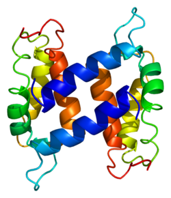
Calcium is second only to oxygen, carbon, hydrogen, and nitrogen in the body, accounting for about 2% of human body weight. Ninety-seven percent of the calcium in the body is found in the bones. Another 1% of calcium is used in the form of ions. The normal concentration of calcium ions is necessary to maintain the integrity of the membrane, the excitability of the muscles, the nervous system and the multiple functions of the cells. The body maintains calcium homeostasis by regulating intestinal calcium absorption, urinary calcium excretion, and bone formation. Calcium-binding protein (CaBP) and its related molecules play an important role in the absorption of calcium and the transmission of calcium metabolism signals. Since calcium homeostasis plays an important role in the health of the body, it is very important to study the mechanism and regulation of calcium-binding proteins for the treatment of related diseases.
The Gene Structure of Calcium-Binding Protein
The cabp gene was isolated from the rat gene pool using the rat CaBP cDNA probe and its structure was confirmed. First, a 30 kb p gene fragment was cloned. Cabp gene is 2.5 kbp long and contains three exons and two introns. The first exon contains only one 5 ' untranslated segment, and the second exon encodes the first calcium binding site and the third exon encodes the second calcium binding site and the 3' untranslated segment. That is, each calcium-binding domain is encoded by a separate exon, respectively. The starting site of transcription was determined by S1 nuclease cleavage map and primer extension method. 31bp from the position of the cap has a TATAAA region, and the upstream of the translation start point is a "CCAAT" box, an Alu-like sequence and (AC)25. The (AG)23 sequences together form the second intron, and the repeats form 3' ends of the upper end of the cap.
The Regulation of Calcium-Binding Proteins and Related Molecules
Studies on calcium-binding proteins are now focused on the regulation of calcium-binding proteins, and several factors are known to regulate calcium-binding proteins. The first is age. As the age increases, intestinal calcium absorption gradually decreases, and intestinal mucosal CaBP also decreases significantly. That is to say, the active transport and passive absorption of intestinal calcium are gradually reduced. The reduction of intestinal calcium absorption in women over 60 years of age is more pronounced than in men. Some people think that because of the lack of estrogen in postmenopausal women, the sensitivity of bone to parathyroid hormone(PTH) is increased, so the secretion of PTH is reduced, so that on the one hand, the increase of blood phosphorus can inhibit the kidney 1,25 (OH)2-D3 (Vitamin D active substitutes, and increase CaBP activity) synthesis; on the other hand, the decrease in PTH secretion also directly reduces the promotion of 1,25 (OH)2-D3 synthesis in the kidney, causing its level to decrease, and thus the absorption of intestinal calcium is also reduced. In rats, the ability of the small intestine to absorb food calcium also gradually decreases with age, and the urinary calcium excretion of the kidney is also age-related, indicating that the concentration of CaBP in the small intestine and kidney is a limiting factor for calcium absorption. To confirm this, the expression of CaBP at different ages was measured, and changes in 1,25 (OH)2-D3 in blood were measured. Experiments showed that the CaBP mRNA in the small intestine decreased significantly in the 2-6 months, and increased significantly in the 13 to 24 months, and the CaBP also decreased in parallel in 2 to 6 months. However, it did not rise with the increase of mRNA; the CaBP in the kidney decreased in 2-13 months, and then the platform was formed, and the situation of CaBP mRNA was similar. These experiments confirmed a decrease in the concentration of CaBP that occurred with age, which may explain the decrease in intestinal calcium absorption and the increase in urinary calcium excretion at this time. The experiment also compared the expression of another CaBP calmodulin. Most scholars believe that the regulation of 1,25 (OH)2-D3 has little effect on the synthesis of calmodulin, but it has also been reported that calmodulin stimulates calcium transport in the lateral membrane of the small intestine in vitro, and 1,25 (OH)2-D3 increases the content of calmodulin in the brush border and is parallel to the calcium transport capacity of these membranes. These results indicate that calmodulin is involved in abnormal changes in age-related calcium transport capacity. The level of CaBP in the small intestine depends on the vitamin D forming active 1,25 (OH)2-D3. In the vitamin D-deficient rats, the concentration of CaBP decreases, after injection of 1,25 (OH)2-D3, CaBP concentration can be restored to normal again. Recent studies have shown that at least 10% of mRNA extracted from rat duodenum directs the synthesis of CaBP, and the content of mRNA directly affects the synthesis of CaBP, thereby affecting the absorption of intestinal calcium, and the levels of 1,25 (OH)2-D3.
References:
- Braunewell K H, Gundelfinger E D. Intracellular neuronal calcium sensor proteins: a family of EF-hand calcium-binding proteins in search of a function. Cell & Tissue Research. 1999, 295(1):1-12.
- Winsky L, Kuåºnicki J. Antibody recognition of calcium-binding proteins depends on their calcium-binding status. Journal of Neurochemistry. 2010, 66(2):764-771.
- Haghighat N G, Ruben L. Purification of novel calcium binding proteins from Trypanosoma brucei: properties of 22-, 24- and 38-kilodalton proteins. Molecular & Biochemical Parasitology. 1992, 51(1):99-110.


